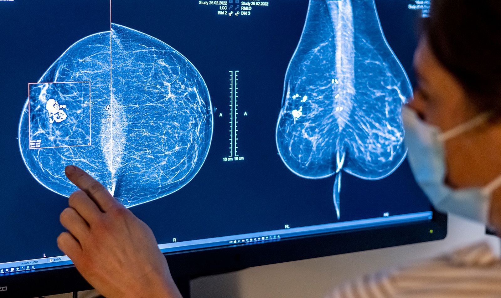
A 38-year-old female with increasing right breast lump since 15
$ 25.99 · 4.9 (112) · In stock

Download scientific diagram | A 38-year-old female with increasing right breast lump since 15 months. Mammogram ( ): An irregular high-density mass with indistinct margins is seen in predominantly upper inner quadrant also extending in the outer quadrant measuring approximately 4.4 × 4.4 × 5.5 cm. Pleomorphic microcalcifications ( ) are seen within the mass, better seen on magnification view. Diffuse trabecular thickening with nipple areolar complex thickening and retraction is seen. Few suspicious right axillary nodes are seen, largest measuring 1.2 × 0.7 cm with 4.5-mm cortical thickness ( ). In view of dense breast parenchyma, further evaluation with CEM was performed to rule out any other lesion in breast, CEM ( ) is suggestive of large unifocal lesion. This is the case of locally advanced breast cancer (stage IIIA), further metastatic work-up was performed. On CT scan, ( ) heterogeneously enhancing mass is seen involving right breast with involvement of overlying skin. Enlarged right axillary, right internal mammary, and right supraclavicular lymph nodes are seen. (CEM, contrast-enhanced mammogram.) from publication: Imaging Recommendations for Diagnosis, Staging, and Management of Breast Cancer | In a rapidly evolving world, with a steep rise in breast cancer incidence, there has been many advances in imaging and therapeutic options of breast cancer care. In this review article, we are trying to cover imaging guideline for cancer detection and their therapeutic | Breast Cancer | ResearchGate, the professional network for scientists.
29-Year-Old With Breast Lump Was Denied Mammogram, Has Stage 4 Cancer

Nita NAIR, Tata Memorial Centre, Mumbai, TMC, Surgical Oncology

Nita NAIR, Tata Memorial Centre, Mumbai, TMC, Surgical Oncology
:max_bytes(150000):strip_icc()/health-conditions-breast-cancer-1500x1000-9de8b63efb5748c493164e369ccd5b27.jpg)
Breast Cancer: Symptoms, Causes, Stages, and Treatment

Meenakshi Thakur's research works Homi Bhabha National Institute, Mumbai and other places

Mammographically detected asymmetries in the era of artificial intelligence, Egyptian Journal of Radiology and Nuclear Medicine

PDF) Imaging Recommendations for Diagnosis, Staging, and Management of Breast Cancer

Breast Lump During Pregnancy: Causes, Types of Lumps & What to Do

Lump In Breast – Cancer Or Benign Cysts In Young Women

A 38-year-old female with increasing right breast lump since 15 months.
Breast density changes over time could be linked to breast cancer risk, study finds