Histology, microscopy, anatomy and disease: Week 3: 2.1
$ 14.99 · 4.9 (272) · In stock

Human alveolar lining fluid from the elderly promotes Mycobacterium tuberculosis intracellular growth and translocation into the cytosol of alveolar epithelial cells - Mucosal Immunology

Lab 3 Histology - The Cell Flashcards
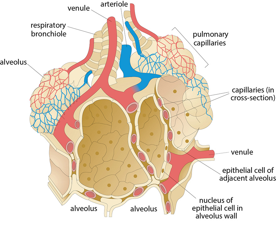
Histology, microscopy, anatomy and disease: Week 3: Figure 2 Schematic diagram of an alveolus, in contact with pulmonary capillaries (Villee, 1989).
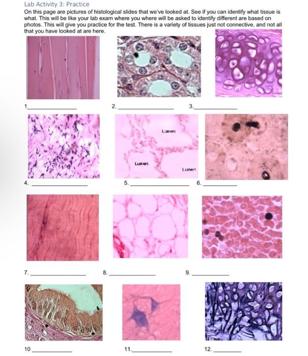
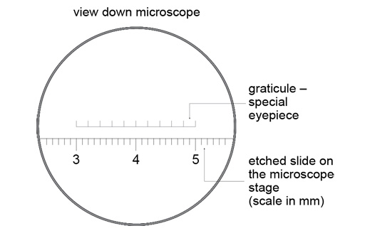
Solved Lab Activity 3: Practice On this page are pictures of

Infection of 3D Brain Organoids with Human Pathogenic Viruses Under Biosafety Level-3 Conditions with Subsequent Inactivation to Study Viral Replication, Pathomechanisms, and Other Viral Infection-Mediated Effects
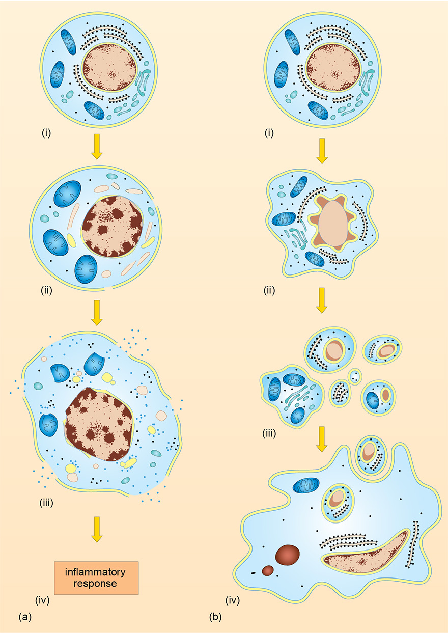
Histology, microscopy, anatomy and disease: Week 4: Figure 5 Schematic diagram comparing (a) necrosis and (b) apoptosis. The first events in necrosis are irregular condensation of the nucleus, swelling of the mitochondria

The spatiotemporal spread of cervical spinal cord contusion injury pathology revealed by 3D in-line phase contrast synchrotron X-ray microtomography - ScienceDirect

Early Diagnosis and Treatment of Cancer by juan jaramillo - Issuu
The Investigation of Caspase-3 and Tumor Necrosis Factor-Alpha Expression in Placentas of Patients with Preterm Premature Rupture of Membranes
Histology, microscopy, anatomy and disease: Week 1: 3

Introduction to Week 1
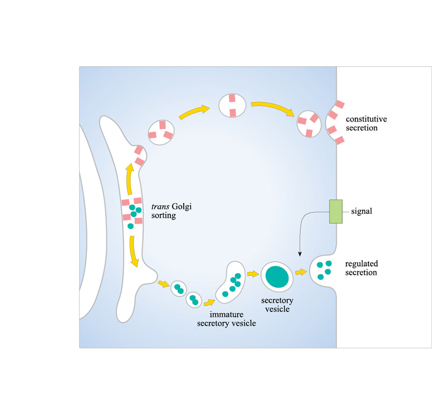
Histology, microscopy, anatomy and disease: Week 3: Figure 1 Secretion of proteins produced in the Golgi apparatus. Secretion may either be constitutive (continuous) or the secreted molecules may be stored in vesicles