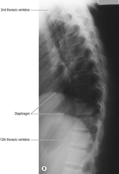
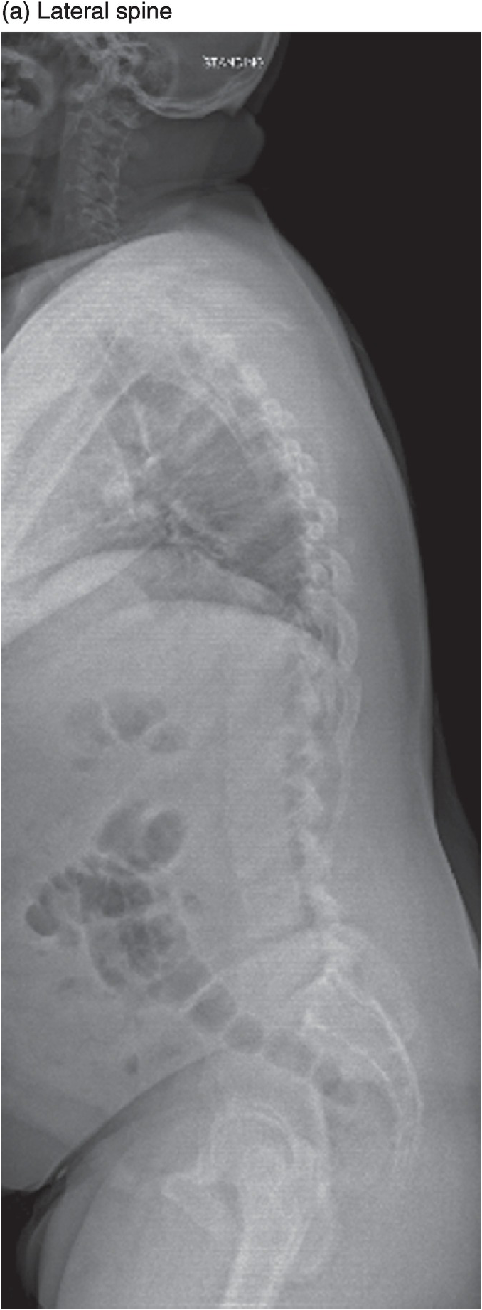
Standing anteroposterior and lateral X-rays of the dorso-lumbar spine
$ 25.50 · 4.6 (144) · In stock

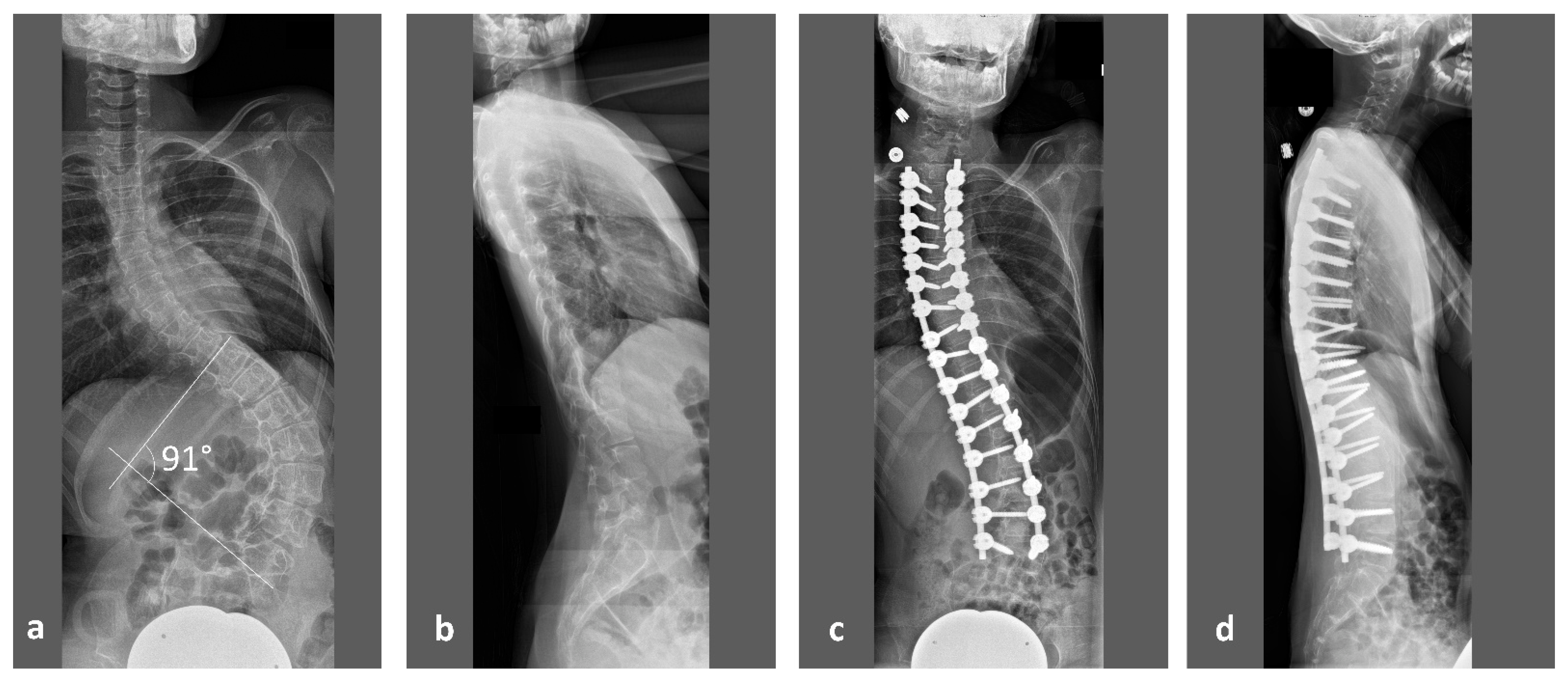
Download scientific diagram | Standing anteroposterior and lateral X-rays of the dorso-lumbar spine showing a failure of the pedicular screws at T11. Note the iatrogenic flat-back deformity with loss of sagittal spine alignment and +ve sagittal vertical axis. from publication: Acute Paraplegia Secondary to Thoracic Disc Herniation of the Adjacent Segment Following Thoracolumbar Fusion and Instrumentation | Proximal junctional disease is a well-recognized postoperative phenomenon in adults who are undergoing long thoracolumbar fusion and instrumentation, and is attributed to increased a junctional stress concentration. In general, the onset of symptoms in these patients is | Paraplegia, Fusion and Segmentation | ResearchGate, the professional network for scientists.
Children, Free Full-Text

Frontiers Case Report: Campylobacter fetus caused pyogenic spondylodiscitis with a presentation of cauda equina syndrome after instrumented lumbar fusion surgery
/wp-content/uploads/2016/03/B97807
Spine clinical cases (Chapter 10) - Postgraduate Orthopaedics

X ray dorso lumbar spine A/P and lateral views showing Kypho-scoliosis

Anteroposterior and lateral view of the dorsolumbar spine showing

Standing anteroposterior and lateral radiographs of the lumbar spine

Lumbar-pelvic-femoral balance on sitting and standing lateral radiographs - ScienceDirect

How to Read a Lumbar X-Ray